
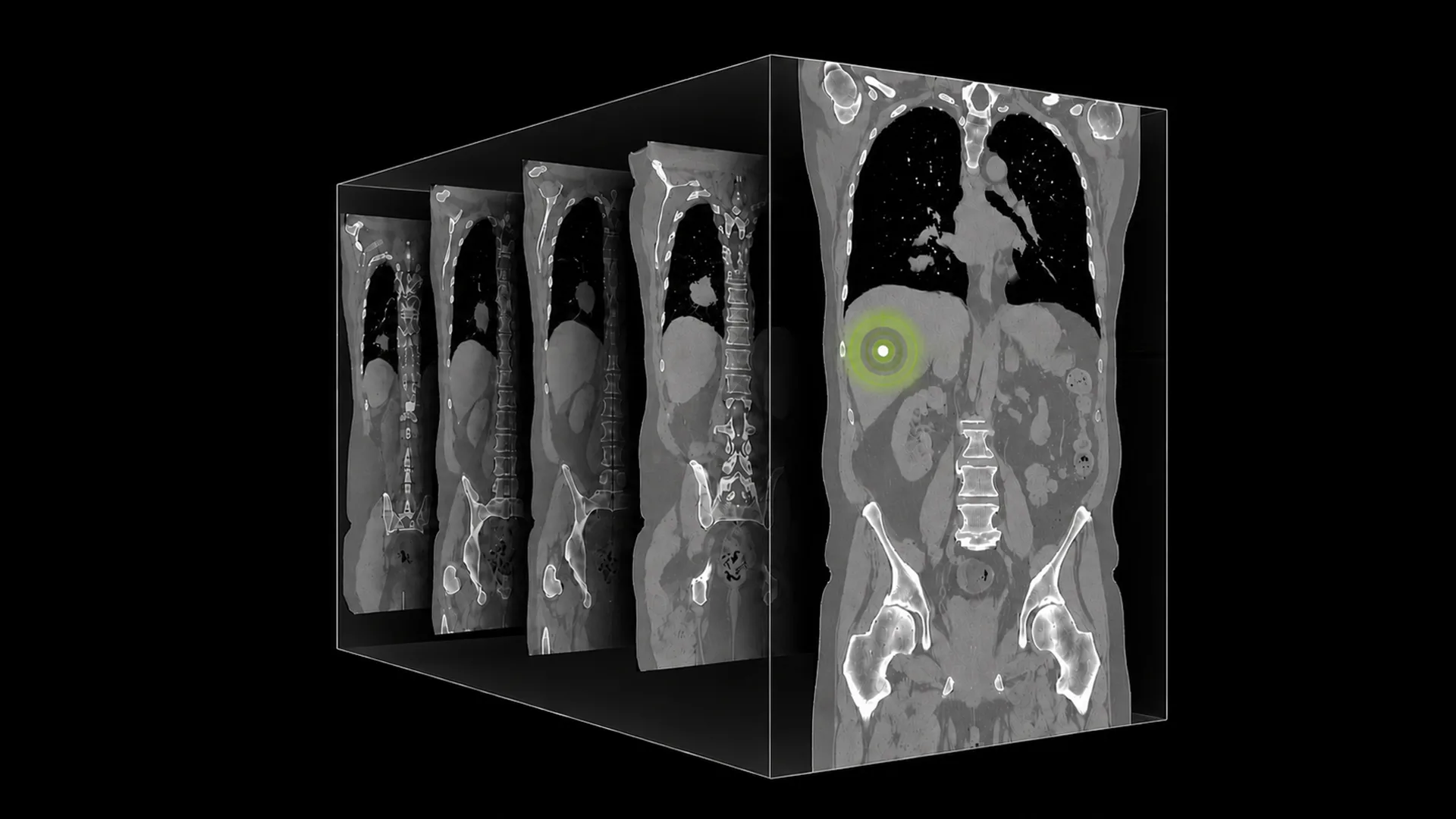
NVIDIA introduced NV-Reason-CT on September 23, 2026, as an open vision language model for analyzing full 3D computed tomography volumes. It generates structured chest and abdominal reports, provides radiologist-style explanations and supports follow-up conversations about findings.
The model extends the approach used in NV-Reason-CXR. A multireader clinical study of that chest X-ray model, accepted at RSNA 2026, found radiologist time savings while maintaining diagnostic accuracy. NV-Reason-CT itself is a research and development foundation for specialized applications—not an autonomous diagnostic system or a cleared clinical product.
Why CT requires native volumetric analysis
An abdominal CT examination can contain 300–600 axial slices. Analyzing those slices independently with a conventional 2D encoder loses relationships that help establish the shape, extent and density of abnormalities across the volume.
NVIDIA identifies three related challenges: preserving three-dimensional anatomy, explaining the evidence behind findings rather than returning labels alone, and supporting iterative questions about differential diagnoses and related anatomical regions. NV-Reason-CT addresses these with a native 3D encoder and a language model trained for structured explanations and multiturn dialogue.
Reports, explanations and follow-up questions
The model’s reporting framework uses a curated ontology of 30 chest and 29 abdominal abnormalities, including lung nodules, pneumothorax, hepatic lesions and renal cysts. Its generated explanations are designed to review anatomical regions systematically, identify relevant findings, discuss alternatives and express uncertainty.
Users can request clarification, investigate a particular finding or ask about a differential diagnosis. The underlying 3D vision transformer preserves continuity between slices, while spatial coordinates accompany the visual tokens into the language model through 3D MRoPE.
Architecture built around a full 3D token grid
NV-Reason-CT pairs the Qwen3.5-4B language model with a 3D vision transformer adapted from Primus and initialized with Colipri weights. All weights are retrained end-to-end on CT data containing structured reports, reasoning traces and internally designed multistep visual question answering.
CT volumes are resampled to 192³ voxels at 2 mm isotropic resolution. Non-overlapping 8×8×8 patches produce a 24×24×24 grid, or 13,824 visual tokens. Rather than merging or reducing them, the architecture passes every token and its 3D coordinates to the language model. Its 3D MRoPE mechanism represents spatial relationships throughout the language-model layers.
Training encourages region-by-region review, consideration of both normal and abnormal findings, calibrated uncertainty and structured conclusions. The intended output is an explanatory assessment rather than a classification label alone.
Two-stage training
Supervised fine-tuning
The first stage combines structured reports, expert radiologist reasoning annotations and general visual question answering. Radiologists supplied detailed CT review dictations describing anatomical checks, significant observations, possible diagnoses and final assessments.
The curriculum contains approximately 550,000 structured question-answer examples covering chest and abdominal anatomy, laterality, localized findings, severity and binary abnormality identification. Refusal examples address invalid prompts and mismatched image-text pairs. Training sources include CT-RATE, NIH CT datasets and CancerVerse.
Synthetic reasoning examples distilled from large language models supplement these materials, using expert annotations as grounding examples.
Reinforcement learning
The second stage applies Group Relative Policy Optimization (GRPO). Rewards reflect the accuracy of identified abnormalities and diagnoses within individual anatomical regions, rather than relying on one global score. This anatomy-aware approach is intended to improve correctness and calibration across chest and abdominal findings after the model has learned the reporting and explanation patterns.
CT-RATE benchmark results
NVIDIA reports that NV-Reason-CT outperforms the published comparison models on CT-RATE classification. Evaluation covered 18 labels with a fixed uniform threshold. NV-Reason-CT answered direct Yes/No prompts without a classification head or task-specific adaptation.
- NV-Reason-CT: native 3D generative VLM; Macro-F1 0.614 and Macro-AUROC 0.871.
- VoxelFM: 3D image-only pretraining; Macro-F1 0.581 and Macro-AUROC 0.870.
- Pillar-0: 3D contrastive model; Macro-F1 0.544 and Macro-AUROC 0.861.
- ClinFusion-8B: fused 2D/3D generative MLLM; Macro-F1 0.442, with Macro-AUROC not reported.
- CT-CLIP: 3D contrastive model; Macro-F1 0.398 and Macro-AUROC 0.733.
- Merlin: 3D contrastive model; Macro-F1 0.358 and Macro-AUROC 0.662.
- MedGemma 1.5: input of up to 85 axial slices; Macro-F1 0.303, with Macro-AUROC not reported.

NVIDIA describes NV-Reason-CT as the first single open model to combine competitive CT classification and report generation. NIH radiologists also reviewed its structured reports and found its generated reasoning clinically plausible. Baris Turkbey, M.D., F.S.A.R., a senior clinician at the National Institutes of Health, highlighted the usefulness of reviewing a systematic explanation alongside the model’s conclusions.
Reporting efficiency and clinical review
NVIDIA says the model can produce detailed structured reports in seconds, allowing clinicians to review and amend the results. Its clinical-impact discussion refers to reports covering 60+ abnormalities, while the specified ontology lists 30 chest and 29 abdominal categories.
The proposed workflow retains radiologist oversight. Reviewers can inspect the generated explanation, compare it with their own assessment and identify disagreements. These reported benefits do not change the model’s status as a research foundation rather than a cleared clinical system.
Research and application development
Researchers can use model checkpoints and post-training recipes to investigate medical-imaging reasoning, adapt the model to institution-specific CT data or incorporate it into multimodal pipelines. NV-Generate-CTMR and NV-Segment-CTMR provide complementary capabilities for synthetic data generation and segmentation.
Potential application areas include radiology workflow software, picture archiving and communication systems, and clinical decision support. Aidoc, HOPPR, Rad AI, Mosaic Clinical Technologies and Raidium are cited as companies operating in relevant markets, not as confirmed adopters.
Example inference workflow
Checkpoints are available through Hugging Face under nvidia/NV-Reason-CT. The GitHub repository provides inference scripts, training configurations and post-training recipes.
The supplied example loads the model with AutoModelForImageTextToText and its processor with AutoProcessor, enables remote code, and uses SDPA attention with torch.float16 on CUDA. A chat template enables thinking, and the processor receives chest_ct.nii.gz through images3d with anatomy_region="chest". Generation is deterministic, uses caching and permits up to 4,096 new tokens before decoding the response.

The illustrated output reviews an IV contrast-enhanced chest CT. It notes a left-sided port catheter ending in the superior vena cava and patent central airways. The principal findings include a large right lower lobe mass with partial collapse, another mass-like lesion in that lobe, and bulky mediastinal and right hilar adenopathy, interpreted as concerning for thoracic malignancy with nodal involvement.
The example also describes scattered bilateral ground-glass opacities with inflammatory, infectious or neoplastic possibilities, lytic lower thoracic spine disease with moderate spinal stenosis, and several indeterminate sclerotic right rib lesions.
Position in NVIDIA’s medical AI portfolio
- NV-Generate-CTMR: generates realistic 3D CT and MRI volumes for research and training-data augmentation.
- NV-Segment-CTMR: segments organs and lesions in 3D CT and MR volumes.
- NV-Reason-CXR: provides reasoning for chest X-ray analysis.
- NV-Reason-CT: provides volumetric CT analysis, structured reports and conversational follow-up.
Together, these models are intended as building blocks for pipelines spanning synthetic data, segmentation and explanatory image analysis. NV-Reason-CT adds chest and abdominal volumetric reporting to that research ecosystem.
Research and product contributors

Andriy Myronenko is a senior research scientist at NVIDIA specializing in computer vision, deep learning and medical image analysis, including 3D MRI/CT organ and tumor segmentation. He collaborates with hospitals including UCSF and Stanford on clinical workflows. He won first place in the 2018 BraTS brain tumor segmentation challenge and earned a PhD from Oregon Health and Science University in 2010, researching non-rigid image and point-set registration.

Supriya V. Thathachary is a senior product manager for Medical AI at NVIDIA and leads MONAI product management. Her work covers medical imaging, multimodal AI research and sensor reconstruction. Her earlier experience includes surgical robotics, ultrasound and software that transformed patient CT scans into segmented virtual models for surgical planning. She holds a Ph.D. in biomedical engineering from the University of Minnesota.

Monty Zarrouk leads product marketing for NVIDIA Medical AI and NVIDIA MONAI. He has two decades of HealthTech product-marketing experience and previously led worldwide healthcare marketing strategy and portfolio positioning at AWS. He holds an MBA from McGill University and a bachelor’s degree in Electrical Engineering from the University of Colorado at Denver.